This article aims to highlight all the most important things to look for when trying to diagnose cranial muscle palsies.
There are 14 cranial nerves in the brain. However, there are only three that supply the eye muscles. They include:
Cranial nerve 3 – Called the Oculomotor Nerve and comes off the brain stem
Cranial nerve 4 – Called the Trochlear Nerve and is the only nerve to come from the back of the brain stem. It’s the longest and thinnest of the nerves.
Cranial nerve 6 – Called the Abducens Nerve and comes out from the brain stem at an awkward angle, making it more susceptible to damage from high intracranial pressure
Third Cranial Nerve – Innervates the majority of the eye muscles including:
- Superior Rectus – elevates the eye
- Lateral Rectus – pulls the eye out
- Inferior Rectus – pulls the eye down
- Inferior Oblique – helps with down gaze and has a torsional (rotational) role.
- Levator muscle – keeps the eye open
NOTE: Parasympathetic nerves which control pupil constriction also run along with the third nerve hence why the pupil may be involved with a third nerve palsy.
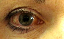
What does a third nerve palsy look like?
- Eye appears down and out
- The patient may have a droopy eye lid
- Pupil may or may not be blown (if pupil blown – red flag, possible aneurysm or tumour)
- The patient complains of side by side/slanted diplopia
Causes of a third nerve palsy:
- Most common is a vascular cause like diabetes or high blood pressure
- Tumour
- Aneurysm (be aware if pupil blown, patient needs to be imaged and treated)
Forth Cranial Nerve –the most confusing nerve! It innervates one muscle only:
- Superior Oblique – Intorts and pulls the back of the eye so the eye looks down. It’s confusing because the muscle action varies depending on which way the eye is looking.
Basic rules:
- If eye is looking inwards towards the nose, the muscle works in an up/down motion
- If eye is looking outwards, the muscle works in a more side to side/twisting motion
What does a fourth nerve palsy look like?
- The eye appears upwards and inwards – known as a ‘nasal upshoot’
- Possible head tilt
- The patient complains of slanted diplopia
- If possible temporal arteritis, the patient complains of a tender scalp, general malaise and jaw claudication.
Cause of a forth nerve palsy:
- Vascular cause like diabetes or high blood pressure
- Tumour
- Congenital (most common cause in the young and often associated with a head tilt that has been long standing)
- Trauma (also quite common due to where the nerve is and because it’s so long and delicate)
- Sometimes temporal arteritis can cause it, especially in an older patient
NOTE: A good way of checking for a fourth nerve palsy is to use the Maddox rod technique where you present the rod (with the lines vertical) in front of the eye that you think is affected. Shine the pen torch and if the eyes are aligned, the patient will see the red line and white spot of the light aligned. If the eye is looking up, the red line appears below the white spot of light.
Sixth Nerve Palsy – controls only one muscle:
- Lateral rectus – pulls the eye out
What does a sixth nerve palsy look like?
- The eye or eyes turns in (cross eyed)
- The patient complains of horizontal (side by side) diplopia.
Causes of six nerve palsy:
- Vascular cause like diabetes of high blood pressure
- Tumour
- High intracranial pressure – be aware of this one and if in a young patient rule out pseudotumour cerebri which can be set off by antibiotic use and recent weight gain (especially in young females)